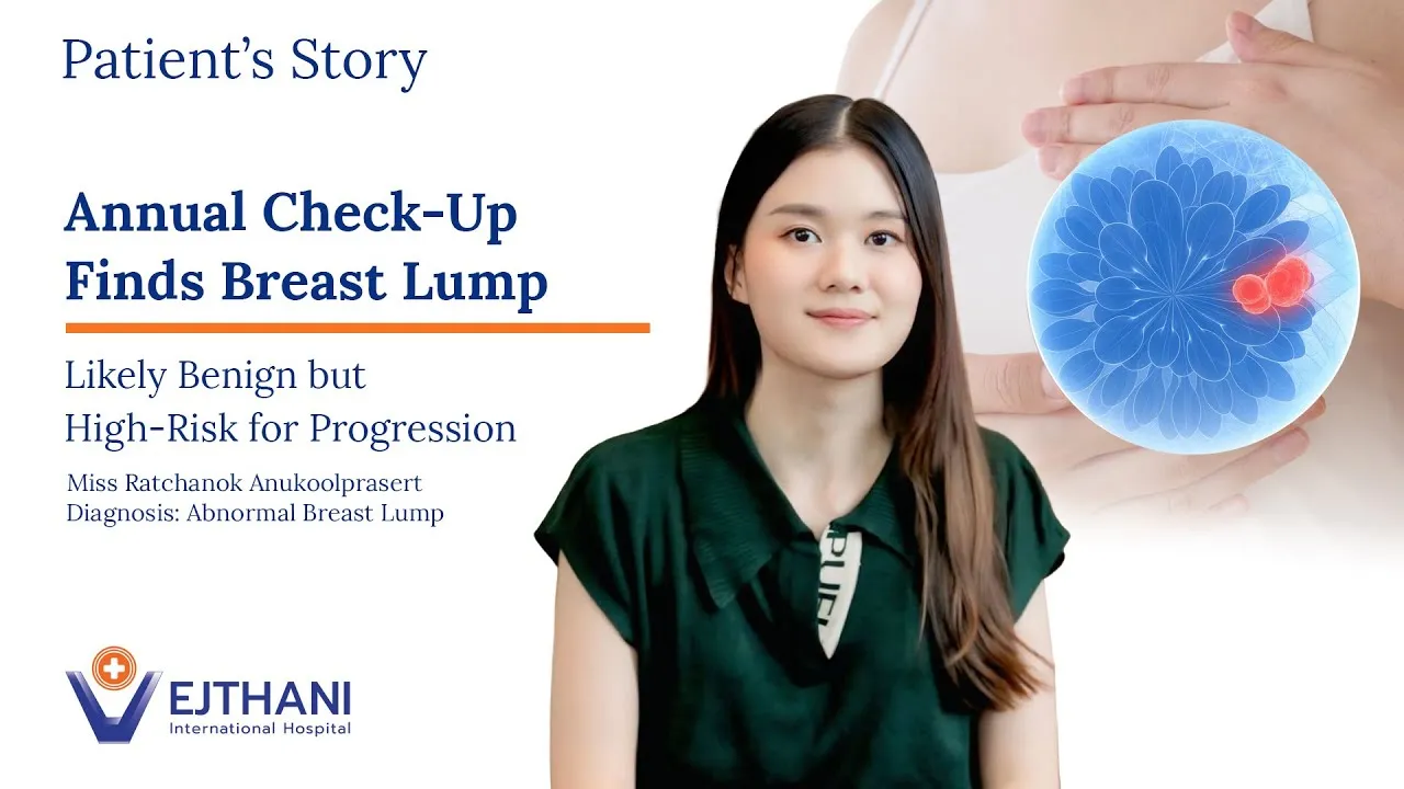
At just 29, Miss Ratchanok went for her very first breast health check—something she had never thought she needed. But that single decision changed everything.
After meeting with Dr. Monchai and undergoing an ultrasound, she learned that there were multiple lumps in her breast. Their characteristics suggested that some carried a higher risk of becoming harmful, which is why a biopsy was performed to determine whether they were benign. Fortunately, the biopsy results confirmed that the lumps were non-cancerous.
Given their potential to progress over time, Dr. Monchai recommended removing them early—before they develop into something more serious.
Her experience is a reminder that breast health checks are essential—even at a young age, and even when you have no symptoms. Early detection makes the difference.
FiLaC (Fistula-tract Laser Closure) is an advanced laser technology designed to treat anal fistula with exceptional precision while preserving sphincter muscle function.
FiLaC delivers controlled laser energy inside the fistula tract to seal it safely—without cutting the sphincter muscle. This minimally invasive approach reduces tissue damage, minimizes postoperative pain, and supports faster recovery, allowing patients to return to daily life sooner.
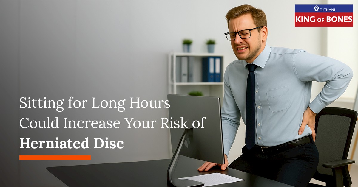
Many people downplay chronic back or neck pain, assuming it is a minor issue that will resolve on its own. However, these symptoms may be an early warning sign of a herniated disc, a condition that, if left untreated, can worsen and significantly impact your daily life.
What is Herniated Disc?
A herniated disc occurs when one of the spinal discs—responsible for absorbing shock and allowing smooth movement—tears or slips out of place. This displacement can press on nearby nerves, causing pain, numbness, or weakness in various parts of the body, such as the back, neck, hips, or legs.
Symptoms of Herniated Disc
Symptoms vary depending on the location of the disc herniation and the degree of nerve compression. Common signs include:
Chronic back or neck pain, especially during movement or heavy lifting
Radiating pain along the nerve path (e.g., down the leg if the disc slips in the lower back, or down the arm if in the neck)
Numbness or weakness in the legs, arms, or hands
Electric shock-like sensations when moving or staying in certain positions
Muscle weakness or limited range of motion
If left untreated, symptoms can worsen over time and may lead to:
Muscle atrophy or increasing weakness
More intense nerve pain and numbness
Loss of bowel or bladder control in severe cases
Causes of Herniated Disc
Age (typically affects those between 30 to 50 years old)
Poor posture or improper use of the spine (e.g., lifting heavy objects incorrectly)
Spinal injury or trauma
Excess body weight or obesity
Lack of regular exercise causes weak back muscles
Diagnosing Herniated Disc
Diagnosis typically begins with a medical history and physical examination. Additional tests may include:
MRI (Magnetic Resonance Imaging): A detailed scan to detect nerve compression and assess the severity of disc damage.
Treatment Options
For mild or early-stage herniation:
Pain relief medications
Physical therapy
Lifestyle adjustments, such as correcting posture and modifying sleeping or lifting habits
For more severe or persistent symptoms:
If the condition does not improve within six months or becomes more severe, such as radiating leg pain, sleep disturbances, difficulty walking, or loss of bladder/bowel control, your doctor may recommend endoscopic discectomy, a minimally invasive spine surgery.
Benefits of Endoscopic Discectomy
Small incision (around 1 cm)
Minimal trauma to surrounding tissue, resulting in less post-operative pain
Faster recovery time and quicker return to daily activities
Preventing Herniated Discs
While age-related degeneration cannot be entirely avoided, you can lower your risk and delay the onset of symptoms by:
Maintaining a healthy weight
Exercising regularly to strengthen core and back muscles
Avoiding smoking
Having a balanced diet
Practicing proper posture when sitting, standing, bending, or lifting
Changing your position regularly when working at a desk
Taking precautions to avoid spine-related injuries
Don’t Ignore Chronic Pain
If you’ve been experiencing persistent back pain, neck pain, or numbness in your limbs, don’t wait for it to become more severe. Consult a spine specialist for an accurate diagnosis and appropriate treatment recommendations.
Knee pain, stiffness, and discomfort during walking can indicate early knee osteoarthritis. Without timely treatment, the condition can progress, causing further cartilage wear, chronic inflammation, walking difficulties, and, eventually, the need for walking aids in daily life.
What is Knee Osteoarthritis?
Knee osteoarthritis occurs when the cartilage covering the joint surface deteriorates, causing the bones within the knee joint to rub against each other. This leads to pain, stiffness, tightness, and reduced mobility. It is common among older adults or individuals who have long subjected their knees to heavy use, such as frequent lifting, running, or climbing stairs.
The condition is increasingly seen in working-age and middle-aged individuals due to lifestyle habits such as prolonged kneeling, sitting on the floor, and excess body weight that place additional stress on the knee joint.
Symptoms of Knee Osteoarthritis
Knee pain, especially when standing, walking, or climbing stairs
Morning stiffness or stiffness after prolonged sitting
Cracking, popping, or grinding sounds in the knee
Swelling or fluid accumulation in the joint
Knee deformity, such as bowlegs or knock-knees
If left untreated, symptoms may worsen over time, affecting mobility, daily activities, and overall quality of life.
Age: Natural decline in the body’s ability to repair cartilage increases the risk.
Gender: Women, particularly those over 50 or post-menopause, have a higher risk than men.
Excess body weight: Adds pressure on the knees, accelerating inflammation and joint degeneration.
Genetics: A family history may increase susceptibility, although not directly inherited.
Improper knee usage: Heavy or repetitive knee movements, frequent stair climbing, prolonged kneeling, squatting, or sitting cross-legged.
Previous knee injuries: Accidents or sports injuries such as meniscus or ligament tears, can increase the risk of early joint degeneration.
Weak supporting structures: Lack of exercise can weaken the muscles and ligaments around the knee, reducing joint stability.
Underlying medical conditions: Rheumatoid arthritis, gout, SLE, and certain blood disorders may also contribute to knee degeneration.
Diagnosis
Your doctor will begin with a detailed medical history, physical examination, and assessment of knee mobility. Imaging studies such as X-ray or MRI may be used to evaluate cartilage damage and surrounding tissues, allowing your specialist to design the most suitable treatment plan.
Treatment Options
Treatment depends on the severity of the condition and may include:
Non-surgical Options
Weight control
Physiotherapy and strengthening exercises
Knee injections (medication or joint lubricants)
Anti-inflammatory medications
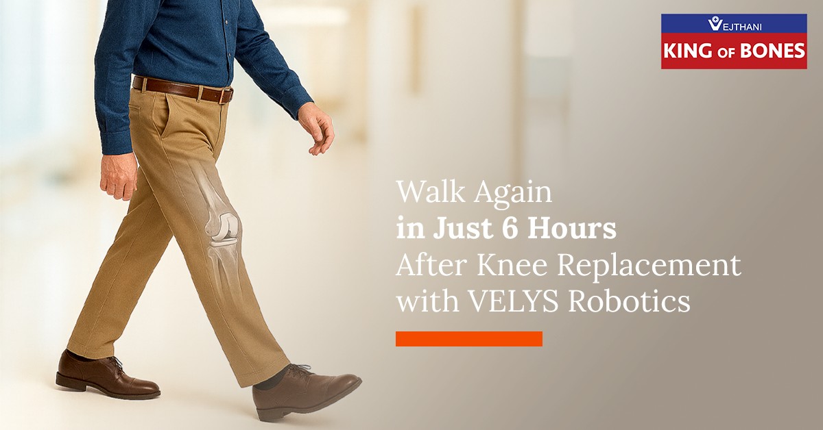
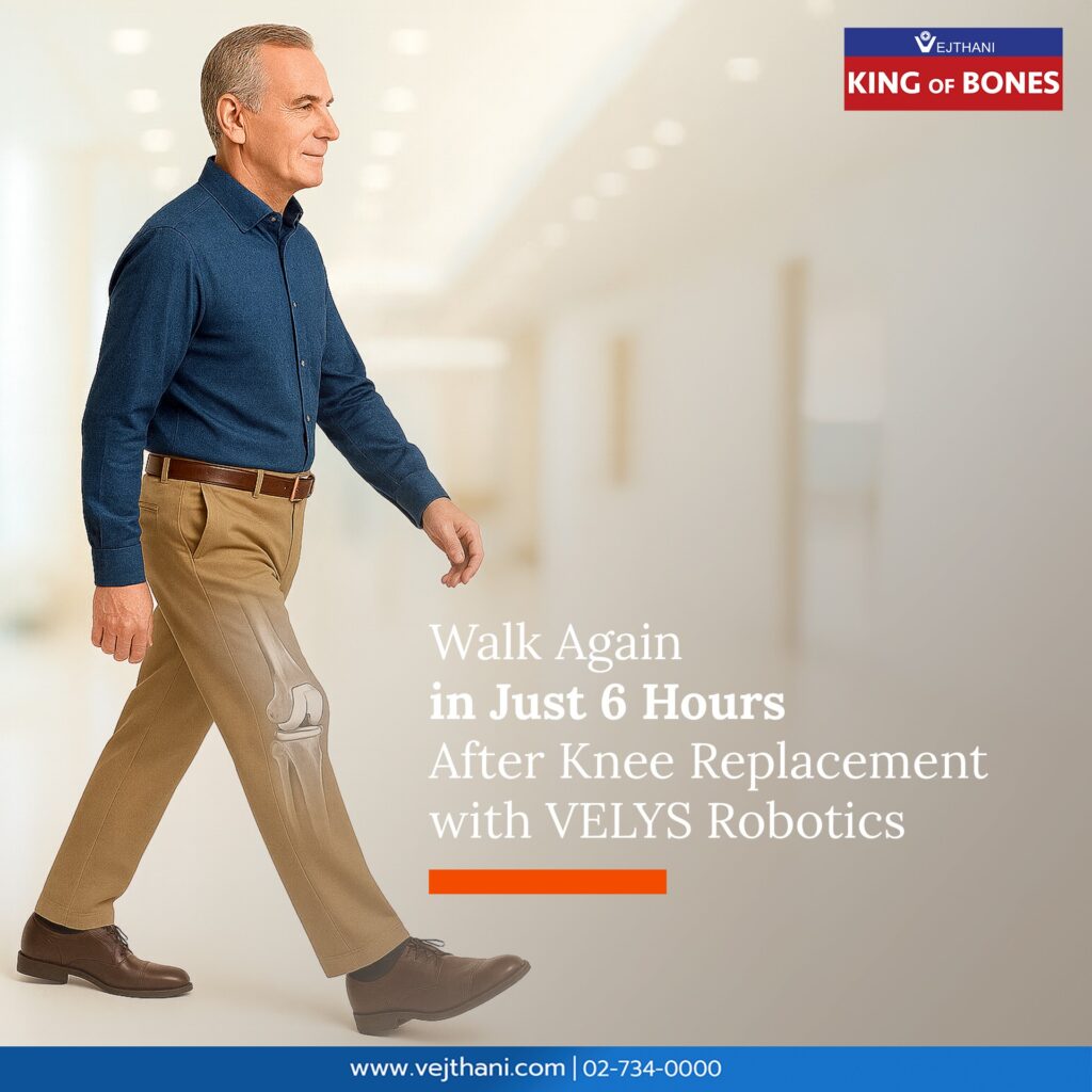
If symptoms are severe and conservative treatments are no longer effective, your doctor may recommend Total Knee Replacement (TKR).
Next-Generation Knee Replacement with the VELYS™ Robotic System
Modern knee replacement is not just about “replacing the joint”, it is about restoring your quality of life.
3D knee imaging for personalized surgical planning
Real-time assessment of leg alignment and joint stability
Reduced impact on surrounding tissues
Faster recovery, less pain, and extended implant durability
Improved movement that feels as natural as possible
Why Personalized Alignment Matters
The VELYS™ system is not a “one-size-fits-all” approach. It uses the patient’s real movement data, walking patterns, knee bending angles, and bone alignment to ensure:
The implant fits seamlessly with the patient’s anatomy
The surgeon achieves optimal precision
Movement feels natural, with faster recovery and confidence in every step
Because VELYS™ does not simply “assist surgery”, it helps you walk againwith your natural movement.
Who is VELYS™ Suitable For?
Ideal candidates include people who:
Have moderate to severe knee osteoarthritis
Have not improved with standard treatments
Want faster recovery and less postoperative pain
Seek a precise and safe surgical option
VELYS™ Robotic-Assisted Solutions
An advancement in knee replacement surgery, enhancing precision, safety, and recovery to restore quality of life.
FAQ: Knee Osteoarthritis
How is knee osteoarthritis treated?
Start with weight management, exercise, and physiotherapy. For advanced cases, robotic-assisted knee replacement with VELYS™ may be recommended for higher precision and faster recovery.
How long is the recovery after VELYS™ robotic knee replacement?
Most patients can walk within 6 hours after surgery and return to everyday life faster.
How is VELYS™ different from conventional knee replacement?
VELYS™ provides real-time 3D imaging, precise implant placement, reduced tissue trauma, less pain, faster recovery, and movement that feels more natural.
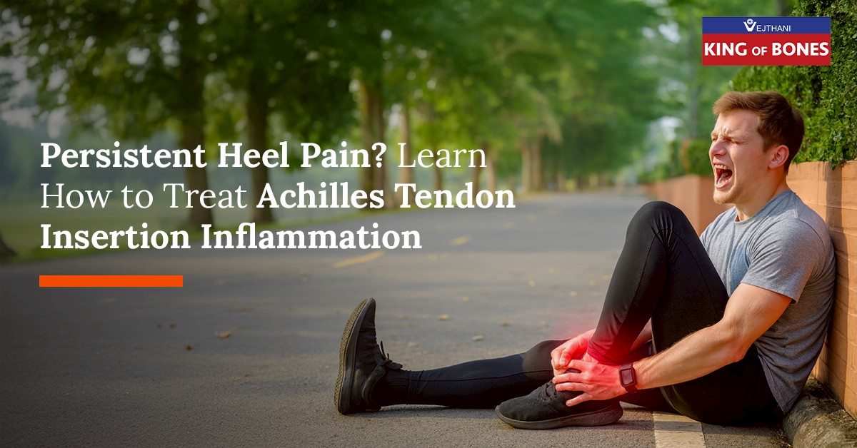
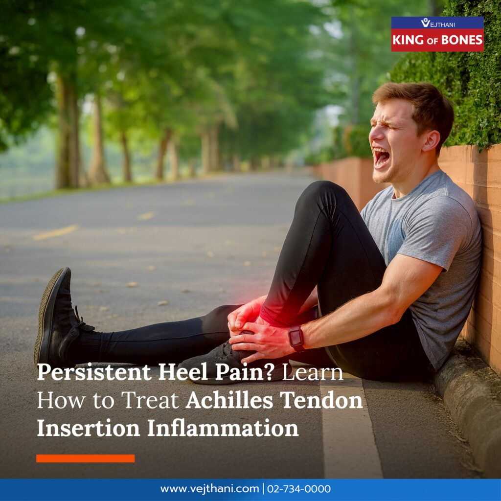
Pain at the back of the heel may not simply be routine soreness. It can be a sign of Insertional Achilles Tendinopathy, which often affects individuals who are engaged in intense exercise, athletes, and those who place repetitive stress on their feet every day. Without proper care, this condition may become chronic or even lead to tendon tears.
What Is Insertional Achilles Tendinopathy?
The Achilles tendon is the largest tendon in the body, connecting the calf muscles to the heel bone. When inflammation occurs at the tendon’s insertion point, it can cause pain, swelling, redness, or tenderness at the back of the heel—especially when standing, walking, or running.
Symptoms of Achilles Tendinitis
Pain at the back of the heel, especially in the morning or after exercise
Swelling and tenderness around the heel
In some cases, heel spurs may develop
Increased pain during running, jumping, or wearing shoes that irritate the heel
Causes of Insertional Achilles Tendinopathy
Repetitive stress such as long-distance running, jumping, or high-impact sports
Wearing improper footwear
Foot abnormalities, such as flat feet or a tight Achilles tendon
Age-related loss of tendon elasticity
Excess body weight, which increases pressure on the heel
Diagnosis
Doctors diagnose this condition through medical history, physical examination, and—if needed—ultrasound or MRI to assess severity and guide a personalised treatment plan.
Treatment Options for Achilles Tendinitis
Non-Surgical Treatment
This is the first-line approach, as most patients improve without surgery. It includes:
Rest and activity modification: Avoid activities that worsen symptoms, such as long-distance running or jumping, giving the tendon time to recover.
Cold compress: Helps reduce inflammation and swelling, especially during flare-ups.
NSAIDs (e.g., Ibuprofen, Naproxen): Reduce pain and inflammation.
Physical therapy: Includes calf stretching and ankle-strengthening exercises.
Supportive devices: Insoles or heel pads help reduce tension at the tendon insertion.
Shockwave therapy: Uses high-energy shockwaves to enhance blood flow and stimulate tendon healing. This technique has become increasingly popular.
Surgical Treatment
Surgery may be considered if symptoms persist for 6–12 months despite non-surgical treatment. Options include:
Debridement: Removing damaged tendon tissue to reduce inflammation and stimulate healing.
Removal of heel spurs: If bone spurs are causing friction at the tendon insertion.
Tendon repair: Recommended when partial tears are present.
Post-operative rehabilitation is essential to restore walking, running, and normal foot function.
Prevention
Warm up and stretch before exercising
Choose well-fitted shoes with good shock absorption
Increase exercise intensity gradually
Maintain a healthy body weight
Regularly stretch the Achilles tendon and calf muscles
Although this condition is not life-threatening, chronic symptoms can significantly affect daily life, making walking or exercising increasingly difficult. If you experience persistent heel pain, it is important to consult a doctor for timely diagnosis and treatment.
For more information, please contact: Orthopedics Center,Vejthani International Hospital Tel. 02-734-0000 Ext. 2298 English Hotline: (+66)85-223-8888
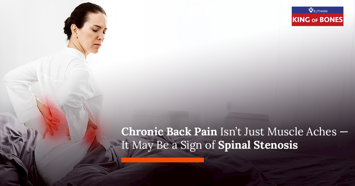
Spinal stenosis is a condition in which the space within the spinal canal — responsible for protecting the spinal cord and nerves — becomes narrowed due to degenerative changes in structures such as the intervertebral discs, ligaments, and joints. This narrowing compresses the nerves, causing pain, numbness, or weakness in various parts of the body, particularly the lower back and legs.
Spinal stenosis is common among older adults and is a major cause of chronic back pain. Without early evaluation and treatment by a spine specialist, it may lead to permanent weakness or loss of mobility.
Causes of Spinal Stenosis
The primary cause is age-related degeneration of the spine, often accompanied by several contributing factors:
Degenerative Disc Disease
Disc shrinkage or bulging puts pressure on the nerve canal.
Facet Joint Hypertrophy
Degeneration leads to bone overgrowth that narrows the canal.
Ligamentum Flavum Thickening
Thickened ligaments reduce the space within the spinal canal.
Spondylolisthesis
Forward slippage of a vertebra narrows the canal and compresses nerves.
Other causes include trauma, previous surgery, or congenital spine abnormalities.
Symptoms of Spinal Stenosis
Symptoms depend on the location and severity of nerve compression. Common symptoms include:
Lower back pain or pain radiating down the legs
Hip or leg pain when walking for extended periods
Worsening pain when leaning backward; relief when bending forward
Numbness, weakness, or heaviness in the legs
Difficulty walking long distances or needing frequent rests
In severe cases: loss of bladder or bowel control — a medical emergency
How is Spinal Stenosis Diagnosed?
Doctors evaluate the condition through history-taking, physical examination, and imaging such as:
X-ray: Shows bone alignment and joint abnormalities
MRI: Provides detailed views of the nerve canal, discs, and nerves
CT Scan: Used when MRI isn’t possible; shows bone structures clearly
Treatment Options
Treatment depends on the severity of the condition and symptoms. Options include:
Non-Surgical Treatment
Suitable for patients with mild to moderate symptoms:
Physical therapy to strengthen back and abdominal muscles
Pain medications or muscle relaxants
Local anti-inflammatory injections
Lifestyle modifications such as avoiding heavy lifting or prolonged back extension
Surgical Treatment
Considered when symptoms are severe or do not improve:
Decompression Surgery: Relieves pressure on the nerves
Spinal Fusion: Stabilizes the spine in cases of vertebral slippage
At Vejthani International Hospital, decompression surgery is performed using microscope or endoscope-assisted techniques, resulting in smaller incisions, less pain, and faster recovery.
Prevention & Self-Care
Regular exercises to strengthen the back and core muscles
Maintain a healthy body weight
Avoid heavy lifting or prolonged sitting in the same posture
Seek early evaluation if chronic back pain or radiating leg pain occurs
Frequently Asked Questions
1. How is spinal stenosis different from a herniated disc?
Both may cause similar symptoms, such as back pain and leg pain. However, a herniated disc occurs when disc material presses directly on a nerve, whereas spinal stenosis results from narrowing of the spinal canal caused by degeneration, often developing gradually and commonly seen in older adults.
2. Does every patient with spinal stenosis need surgery?
No. Most patients improve with non-surgical treatments such as physical therapy, injections, and lifestyle adjustments. Surgery is recommended only for those with progressive numbness, weakness, limited walking ability, or loss of bladder/bowel control.
3. How long is the recovery after spinal stenosis surgery?
With modern minimally invasive techniques, most patients can walk within 1–2 days after surgery and return to everyday life within a few weeks. Recovery time depends on individual health and rehabilitation progress.
4. What types of exercise are safe?
Choose low-impact exercises that strengthen the back, core, and legs, such as swimming, water walking, gentle yoga, or senior-friendly Pilates. These help reduce spinal pressure and prevent recurrence.
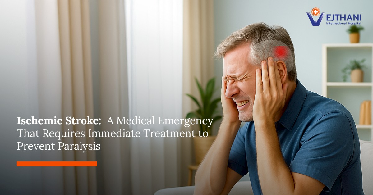
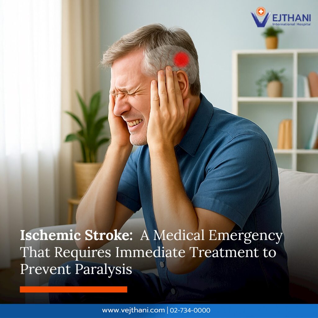
Ischemic stroke occurs when blood flow to the brain is blocked or severely reduced, depriving the brain of oxygen and essential nutrients. When this happens, brain cells begin to die within minutes. Stroke is one of the leading causes of death and long-term disability, especially if left untreated.
Causes of Ischemic Stroke
An ischemic stroke happens from a blockage in the brain’s blood vessels or in the arteries supplying blood to the brain. Common causes include:
Blood clots that form in the brain’s arteries or travel from the heart to block cerebral vessels
Atherosclerosis, where fatty deposits narrow blood vessels
Atrial fibrillation, which increases the risk of clot formation in the heart
Underlying health conditions, such as diabetes, hypertension, and high cholesterol
Lifestyle factors like smoking, excessive alcohol consumption, and lack of exercise
Symptoms of Ischemic Stroke
Symptoms usually appear suddenly and require urgent medical attention. The BEFAST rule helps identify early warning signs:
B – Balance: Sudden loss of balance, dizziness, or unsteady walking
E – Eyes: Blurred vision, double vision, or vision loss in one or both eyes
F – Face Drooping: One side of the face appears uneven or cannot smile symmetrically
A – Arm Weakness: Weakness or numbness in one arm or leg
S – Speech Difficulty: Slurred speech, inability to speak, or difficulty understanding speech
T – Time to Call: Call an ambulance immediately if any of these signs appear
Other possible symptoms include severe dizziness, double vision, loss of coordination, or loss of consciousness.
Diagnosis of Ischemic Stroke
Doctors use several tests to identify the cause and location of the blockage:
Neurological and physical examinations
CT Scan or MRI
MRA/CTA to examine cerebral blood vessels
ECG to detect abnormal heart rhythms
Blood tests to assess risk factors
Treatment for Ischemic Stroke
Treatment must be initiated urgently to reduce brain damage.
Intravenous thrombolytic therapy (rt-PA) within 4.5 hours of symptom onset
Medication to prevent recurrent clots, such as antiplatelets or anticoagulants
Supportive care, including blood pressure control, glucose management, and rehabilitation
Mechanical thrombectomy is a highly effective method to remove clots blocking brain arteries. A neurovascular specialist inserts a small catheter through an artery in the groin and retrieves the clot from the affected brain vessel. Using Biplane Digital Subtraction Angiography (Biplane DSA) enables specialists to view real-time images of the brain’s blood vessels from two angles simultaneously. This enhances precision and reduces procedural risk.
Vejthani International Hospital’s neurovascular team is equipped to perform thrombectomy using advanced Biplane DSA technology. This imaging system improves treatment accuracy, minimizes complications, and ensures holistic, comprehensive care for every patient.
Benefits of Thrombectomy with Biplane DSA
Reduces brain damage and increases chances of recovery
Lowers the risk of additional tissue injury
Shortens treatment time and increases survival rates
Suitable for patients who arrive within 24 hours of symptom onset
Prevention of Ischemic Stroke
Maintain healthy blood pressure, glucose, and cholesterol levels
Eat a balanced diet with reduced salt and saturated fat
Exercise regularly
Quit smoking and limit alcohol consumption
Undergo annual health check-ups to detect early risks
Ischemic stroke is a race against time. Recognizing symptoms early and seeking immediate treatment can significantly improve survival outcomes and reduce long-term disability. With today’s advanced Biplane DSA technology, treatment is safer and faster.
Frequently Asked Questions
Can Ischemic Stroke be treated?
Yes. If treated promptly, especially within 24 hours using Biplane DSA—patients have a significantly higher chance of recovery and reduced disability.
How is Biplane DSA different from conventional treatment?
Biplane DSA provides simultaneous two-angle imaging of cerebral blood vessels, giving doctors clearer visualization of the clot’s location. This increases precision and enhances safety during the procedure.
What should I do when stroke symptoms appear?
Call an ambulance or go to the hospital immediately. Every minute counts, as brain cells begin to die within minutes of symptom onset.
We use cookies to manage your personal information in order to provide you with the best personalized user experience on our website. If you continue using the website, we assume that you accept all cookies on the website. Find out more.