Brain Aneurysm Treatment
Overview
Brain aneurysm is a potentially life-threatening condition that demands immediate care and treatment. Vejthani Hospital provides expert treatment of brain aneurysms in Thailand.
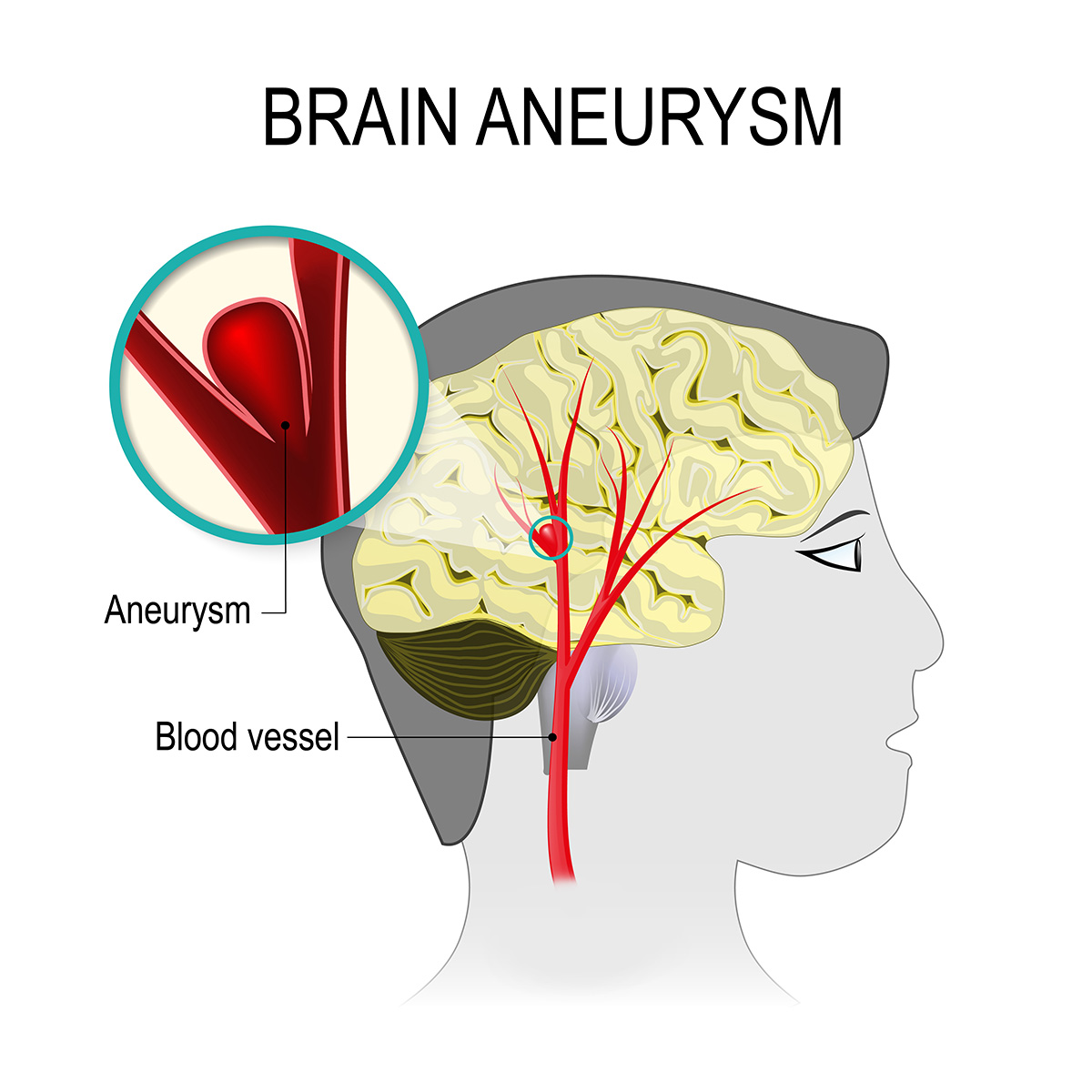
Brain aneurysms, also known as cerebral aneurysms, is a bulge or ballooning in a blood vessel in the brain, filled with blood. Brain aneurysms develop as a result of thinning artery walls. Because those areas of the vessels are weaker, aneurysms frequently form at forks or branches in arteries.
Brain aneurysms show no symptoms until they rupture or leak, causing bleeding in the brain, known as a hemorrhagic stroke. Hemorrhagic stroke could lead to brain damage and is life-threatening. A ruptured brain aneurysm typically develops between the brain and the delicate tissues that cover the brain; this is called subarachnoid hemorrhage.
However, there are brain aneurysms that do not rupture or create any symptoms or problems. Aneurysms are commonly found during examinations for other disorders. A variety of techniques are used as treatments for ruptured aneurysm, aim to block blood from entering into the aneurysm and to divert blood flow across the aneurysm to prevent rupture.
Symptoms
- Ruptured aneurysm: The primary sign of an aneurysm rupture is an abrupt, severe headache. The following are typical warning signs and symptoms of an aneurysm rupture in addition to a severe headache:
- Nausea and vomiting
- Blurred vision
- Sensitive to light
- Stiff neck
- Confusion
- Seizure
- Loss of consciousness
- Leak aneurysm: An aneurysm may occasionally leak small amount of blood which will result in an abrupt, excruciating headache. Leaking is frequently followed by a more serious rupture.
- Unruptured aneurysm: There could be no symptoms if a brain aneurysm does not rupture. However, a larger, unruptured aneurysm could put pressure on the brain’s tissues and nerves that, potentially will lead to:
- Facial numbness on one side
- Pain above and behind one eye
- Dilatation of pupil
- Vision change or double vision
Seek immediate medical attention if you are with someone who has a sudden, severe headache, loses consciousness, or has a seizure.
Cause
The causes of brain aneurysms are still unknown, or the causes of leakage or rupture, but anything that increases your blood pressure can be dangerous. Higher blood pressure makes blood push harder against blood vessel walls, which may increase the risk.
Risk factors
The likelihood of a brain aneurysm or an aneurysm rupture can increase because of artery wall weakness caused by the following:
- Age: Adults are more prone to brain aneurysms than children.
- Gender: It affects women more frequently than men.
- Family history: Family members such as parents, siblings, or children that have a history of brain aneurysms.
- Genetic disorders: such as Ehlers-Danlos syndrome, Marfan syndrome, neurofibromatosis type 1, or Loeys-Dietz syndrome, are connective tissue genetic disorders.
- Other diseases: Having an uncommon blood vessel condition such as cerebral arteritis, fibromuscular dysplasia, or arterial dissection. Also, people suffering from polycystic kidney disease.
If you are within one of the high-risk categories for aneurysms, consult with a doctor at Vejthani Hospital to discuss preventive measures and treatment options to reduce your risk.
Diagnosis
The doctor will evaluate whether there has been bleeding into the area between the brain and surrounding tissues if the patient suddenly develops a severe headache or other symptoms that could be caused by a ruptured aneurysm.
In addition, if the patient is experiencing signs of an unruptured brain aneurysm, such as double vision, pain behind the eye, or vision alterations, they can be subjected to additional testing.
Diagnostic tests consist of the following:
- Imaging tests:
- Computerized tomography (CT): utilize a specific X-ray; this test is used to evaluate whether the patient has a brain hemorrhage or another kind of stroke. As part of the test, a dye maybe be injected that facilitates the visualization of blood flow in the brain and reveal the presence of an aneurysm.
- Magnetic resonance imaging (MRI): To produce precise images of the brain in either 2D or 3D, this imaging method combines radio waves and a magnetic field. The existence of an aneurysm may be discovered via MR angiography, a type of MRI that carefully examines the arteries.
- Cerebrospinal fluid test: The process of using a needle to extract cerebrospinal fluid from your spine is known as a lumbar puncture. Red blood cells will most likely be present in the fluid surrounding the brain and spine (cerebrospinal fluid) if the patient experienced a subarachnoid hemorrhage. A test of their cerebrospinal fluid can help make a diagnosis if they have symptoms of a ruptured aneurysm.
- Cerebral angiogram (Cerebral arteriogram): This treatment involves inserting a thin, flexible tube (catheter) into a major artery, typically in the wrist or groin. To reach the arteries of the brain, the catheter passes through the heart. The brain’s arteries are shown by a particular dye that is injected into the catheter. The condition of the arteries can then be seen in detail and an aneurysm can be found using a series of X-ray scans. When other diagnostic tests are insufficient, a cerebral angiography is commonly done.
- Screening for brain aneurysm: It is not advised to use imaging examinations to check for unruptured brain aneurysms unless the patient is at a high risk. Discuss the advantages of a screening test with health care practitioner.
Treatment
Surgery
- Brain aneurysm surgery: A ruptured brain aneurysm can usually be treated by the following:
- Surgical clipping is a procedure to close off an aneurysm: To access the aneurysm and find the blood supply that feeds the aneurysm, the neurosurgeon must remove a portion of the skull then a tiny metal clip is then inserted on the aneurysm’s neck by the neurosurgeon to halt blood flow to it.
- Endovascular treatment: compared to surgical clipping, is a less invasive process. To reach the aneurysm, the surgeon threads a catheter into your body from an artery, typically in your wrist or groin. The aneurysm is then removed from inside the blood vessel using a flow diverter, an intraluminal flow disrupter, a stent, or coils, or various combinations of these devices.
- Flow diverters: Tubular stent-like implants (flow diverters), which act by diverting blood flow away from an aneurysm sac, are among the more recent therapies for brain aneurysms. The diversion blocks blood flow inside the aneurysm and encourages the parent artery to be rebuilt. Larger aneurysms that cannot be safely treated by other methods may benefit especially from flow diverters.
Other treatments for ruptured aneurysms
It is the goal of additional treatments to treat ruptured brain aneurysms and manage symptoms or complications.
- Medications:
- Pain relievers: acetaminophen for pain, is a potential treatment for headache pain.
- Calcium channel blockers: might reduce the possibility of experiencing severe symptoms from vasospasm, which is a potentially dangerous
complication of a ruptured aneurysm. Nimodipine lowers the risk of delayed brain injury brought on by insufficient blood flow after subarachnoid hemorrhage.
- Anti-seizure medications: could be applied to treat aneurysm-related seizures. These drugs include valproic acid, levetiracetam, phenytoin, and others.
- Interventions to avoid stroke from insufficient blood flow: IV injections are used as interventions to dilate the blood vessels that raises the blood pressure to counter those resistances of narrowed blood vessels.
Angioplasty is a substitute treatment for stroke prevention. In this treatment, a surgeon inserts a catheter to widen a cerebral blood artery that has become restricted due to vasospasm. To dilate blood arteries in the affected area, a medication known as a vasodilator may also be administered. - Ventricular or lumbar draining catheters and shunt surgery: It is possible to insert a catheter to drain extra fluid into an external bag from the areas inside the brain that are filled with fluid or from the region around the brain and spinal cord. It can reduce pressure on the brain brought on by hydrocephalus brought on by a ruptured aneurysm.
- Rehabilitative therapy: Physical, linguistic, and occupational therapy may be required to relearn skills after brain damage following a subarachnoid hemorrhage.
Treatment for unruptured brain aneurysms
- Endovascular coiling: An unruptured brain aneurysm can be closed off using a surgical clip, an endovascular coil, or a flow diverter to help stop it from rupturing in the future. The potential risks may outweigh the any possible benefit.
When providing therapy suggestions, one should take into consideration the following:
- The size, location, degree of irregularity, and general appearance of the aneurysm.
- Patient’s age and general health.
- Family history of ruptured aneurysm
- Congenital issues that increase the risk of an aneurysm rupturing
Consult with the doctor about taking medication to control the high blood pressure. Controlling the blood pressure well may reduce the chance of rupture if the patient has a brain aneurysm.