
Hemorrhoid Grades 1–4: Why Surgery Is Not Always the Answer
Internal hemorrhoids are graded into 4 different categories according to the severity of the disease. Grade 1 The hemorrhoids or the swollen veins bulge inside

Select Topic
If your doctor has recommended a TEE test (transesophageal echocardiography), you may be wondering why it is needed. Most heart conditions are initially diagnosed and assessed by TTE or transthoracic echocardiography. Transesophageal echocardiography is used selectively when more detailed anatomical information is needed, when TTE findings are unclear, or when planning and guiding treatment.
A TEE test (Transesophageal Echocardiography) is a specialized ultrasound that helps doctors examine the heart in greater detail. During the procedure, the cardiologist gently passes a thin, flexible tube with a small ultrasound probe through the esophagus, which sits directly behind the heart. This position helps capture clear, detailed images without interference from the ribs or lungs.
A TEE heart test can provide clearer information about the heart valves, heart chambers, major blood vessels, and blood flow. It may also help identify blood clots, heart infections, congenital heart defects, or other structural abnormalities that need further evaluation. Although TEE test can provide highly detailed heart images, doctors do not use it for routine screening. Instead, cardiologists usually recommend it when they need more information to confirm a diagnosis or guide treatment.
Both a Transesophageal Echocardiography (TEE test) and a transthoracic echocardiogram (TTE) use ultrasound to examine the heart. Each test, however, serves a distinct purpose. Doctors typically start with a TTE, as it’s non-invasive. If more detailed imaging or a closer look at specific heart structures is needed, they may then recommend a TEE.
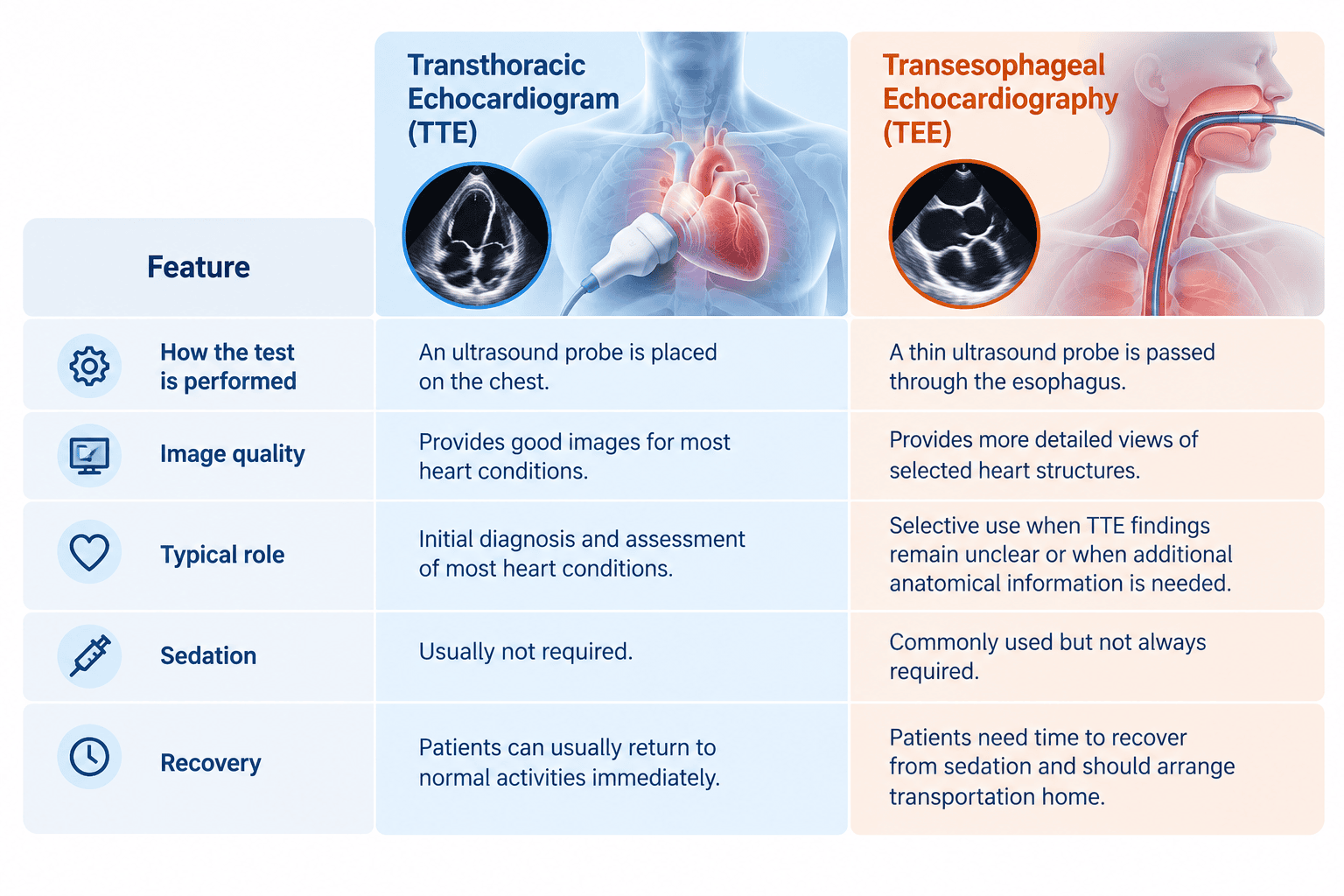
Cardiologists use Transesophageal Echocardiography selectively to answer important clinical questions, clarify uncertain findings, and support treatment planning.
A cardiologist may use this examination to:
Before recommending TEE, the cardiologist considers the patient’s symptoms, medical history, previous test results, and the specific information needed for diagnosis or treatment.
Transesophageal Echocardiography provides detailed anatomical views of selected heart structures, helping cardiologists with diagnosis, clinical decision-making, and certain cardiac procedures.
Cardiologists may also use TEE to guide structural heart procedures, support certain heart surgeries, and assess treatment results after an intervention.
According to Dr. Prayuth Rasmeehirun, cardiologist specializing in cardiac imaging at Vejthani International Hospital, the decision to perform a TEE test depends on each patient’s symptoms, medical history, and previous test results. Cardiologists carefully evaluate all available information before selecting the most appropriate examination to obtain the detail needed for accurate diagnosis and treatment planning.
Preparing for a TEE test usually involves a few simple but important steps. These steps help the examination go smoothly, reduce potential complications, and enable doctors to obtain clear images of the heart.
Before the procedure, patients should:
During a TEE test, patients can expect several steps designed to keep them comfortable while the cardiologist examines the heart.
The nurse checks the patient’s vital signs, helps the patient get into a comfortable position, and monitors heart rate, blood pressure, and blood oxygen levels. The nurse also places an intravenous (IV) line if medications are needed during the procedure. Next, the doctor sprays a local anesthetic into the throat. For some patients, the doctor may also administer conscious sedation to help them relax and reduce discomfort during the examination. Patients continue to breathe normally throughout the procedure.
The patient lies on the left side, and the nurse places a mouth guard to protect both the teeth and the ultrasound probe. The cardiologist then gently guides the ultrasound probe through the mouth and into the esophagus. Throughout the procedure, patients continue to breathe normally.
The cardiologist examines the heart from multiple angles and evaluates its structure and function. The examination also includes capturing detailed images of the heart valves, chambers, blood flow, and nearby blood vessels to support diagnosis and treatment planning.
After completing the examination, the cardiologist carefully removes the ultrasound probe. The medical team then monitors the patient for a short period before allowing them to go home or continue with the planned treatment. The examination itself usually takes about 30–45 minutes, while the entire visit, including preparation and recovery, typically takes 1–2 hours.
Many patients worry that a TEE test will be painful. In most cases, the examination causes only temporary discomfort rather than significant pain. A local anesthetic numbs the throat, and sedation helps patients stay relaxed throughout the procedure.
During and after the examination, patients may experience:
Although most patients recover without any problems, they should contact their doctor if they experience severe chest pain, difficulty swallowing, persistent bleeding, fever, or trouble breathing after returning home.
A TEE heart test is generally safe. However, like other medical procedures, it may cause some side effects or complications, although serious problems rarely occur.
Possible side effects and risks may include:
Patients should contact a doctor immediately if they experience severe chest pain, difficulty swallowing, persistent bleeding, fever, trouble breathing, or worsening symptoms after returning home.
After a TEE test, most patients can go home and rest on the same day. To support a safe recovery, patients should follow a few simple aftercare instructions:
If a doctor has recommended a TEE test or if previous heart test results need further evaluation, speaking with a cardiologist can help clarify the next step. This examination is not a routine screening test, so a cardiologist will consider each patient’s symptoms, medical history, and test results before deciding whether it is appropriate.
At Vejthani International Hospital, our Cardiac Center provides comprehensive heart evaluations using advanced cardiac imaging technology, including TEE testing when clinically appropriate. Our cardiologists focus on accurate diagnosis, individualized treatment planning, and patient-centered care at every step.
A TEE test helps cardiologists see certain heart structures in greater detail. It can support diagnosis, guide treatment planning, and help evaluate conditions such as heart valve disease, blood clots, infections, or structural heart abnormalities when more information is needed.
A TEE test usually takes about 30-45 minutes. Patients may need additional time before and after the examination for preparation, sedation, monitoring, and recovery.
Common TEE heart test side effects may include a mild sore throat, throat irritation, drowsiness, or fatigue from sedation. Serious complications are rare, but patients should contact a doctor if they develop severe chest pain, difficulty swallowing, bleeding, fever, or trouble breathing.
TEE stands for Transesophageal Echocardiography. It is a specialized ultrasound examination that allows cardiologists to view the heart from the esophagus, which sits directly behind the heart.
During a TEE procedure, a cardiologist gently guides a thin ultrasound probe through the mouth and into the esophagus. The probe then captures detailed images of the heart valves, chambers, blood flow, and nearby blood vessels.
For more information, please contact
Cardiac Center, Vejthani International Hospital
Call: (+66)2-734-0000 Ext. 5300
English Hotline: (+66)85-223-8888