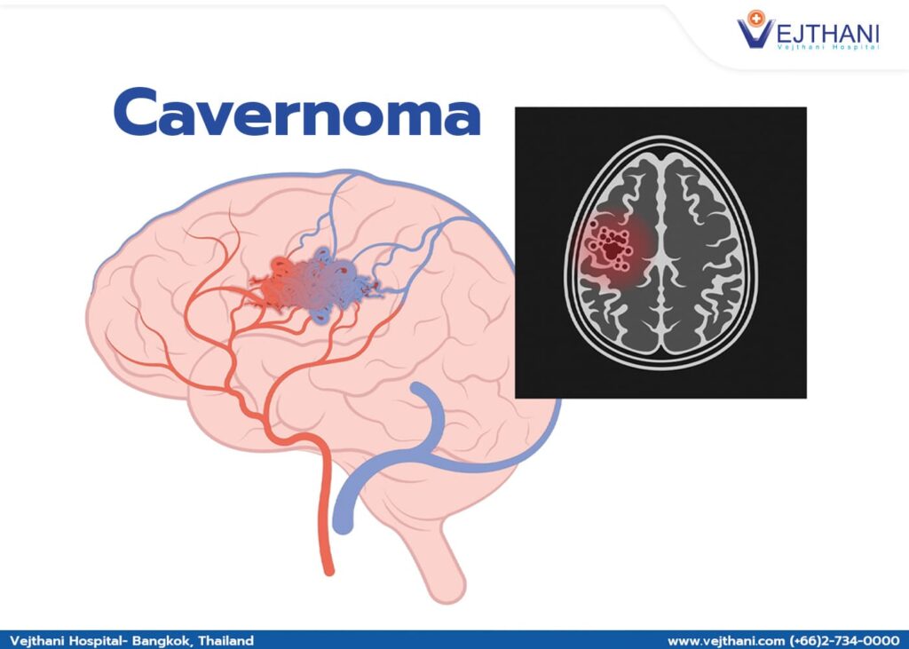
A cavernous bleeding or cavernous hemangioma or cavernoma, or cerebral cavernous malformation is an abnormal collection of tiny capillaries. A cavernous hemangioma can occur from the bleeding of the thin wall of the capillaries because the blood flow within these capillaries has slow or no movement at all. The brain or brain stem is the most crucial body part for this disease. But sometimes, the spine and other body parts may be involved. Bleeding in the brain can cause seizures or stroke.
What does a Cavernous Hemangioma look like?
The hemangioma size ranges from inch to dime-size or larger, filled with blood and separated by connective tissue. Cavernous hemangiomascan affect 1 in 200 people, but mostly 1 in 10 patients suffer from the disease.
How do you know if you have cerebral cavernous hemangioma?
Only 1 in 10 people can have symptoms like seizure or stroke; most patients detect this disease after a brain scan. Some patients with rupture hemangioma may show no signs at all.
There is a chance to have more than one cavernous hemangioma because malformation can occur anywhere and remain stable for many years, even if there is no symptom. A single hemangioma may cause internal bleeding and brain damage while the signs appear.
Twenty percent of cavernous hemangioma is hereditary because of gene mutation. If parents have a history of having cerebral cavernous, there is a 50% chance that their children will develop the disease.
Arteriovenous malformation (AVM) is another type of malformation. They are similar in size and can cause rupture, but the difference between AVM and cavernoma is how the blood flows inside the capillaries. AVMs have a much higher pressure, and it is more dangerous.
Capillary telangiectasias is also a type of blood vessel malformation that rarely causes rupture and damage to the brain.
Symptoms
The symptoms depend on size, the number of hemangiomas, where they are present, and their severity in different individuals.
The most common initial symptom is seizures
Unstable gait or balance
Dizziness, impaired hearing, headache
Muscle weakness in the arms and legs
Impaired vision (Blurred vision, double vision, loss of vision)
Impaired speech
Impaired memory
What causes Cerebral Cavernous Hemangiomas?
There is no specific research on the cause of this hemangioma.
Some research found that this abnormality occurred due to the developmental venous anomaly (DVA), and patients who took Radiation treatment to the brain or spine can also develop cavernoma.
What are the complications of having Cavernous Hemangiomas?
The primary complications that may arise are seizures or stroke. Post-operative complications could be strokes, paralysis, coma, or death. Several factors influence the level of risk, including the overall well-being, the size and location of the lesion, and the volume of lesions.
Diagnosis
Imaging test: magnetic resonance imaging (MRI), susceptibility-weighted imaging is a special type of scan. It can detect the tendency of bleeding or swelling around the lesion and can predict the risk of further complications.
Treatment
How is Cerebral Cavernous Hemangioma treated?
The treatment approach is determined by the lesion’s location and the uncontrolled symptoms. In cases where no symptoms are present, the doctor may wait to oversee brain scans or prescribe medications. However, if symptoms persist, especially neurological conditions or those unresponsive to medicine, and the presence of a hemangioma is confirmed, surgical intervention becomes the primary and preferred treatment method.
Prevention
In most cases, it is impossible to prevent cavernous malformations because of the nature of malformation we cannot predict. But if there is a high chance of a genetic condition, pregenetic testing can be done. Not modifying daily lifestyle can cause a high probability of hemorrhage. Ensure your blood pressure, cholesterol, and glucose level are within the range. Some medications like aspirin and ibuprofen can cause thin blood vessels. They may worsen the bleeding if they are present in your body during malformations.
We use cookies to manage your personal information in order to provide you with the best personalized user experience on our website. If you continue using the website, we assume that you accept all cookies on the website. Find out more.