


Breast cancer is the most prevalent cancer among women worldwide, occurring in approximately 1 in 8 women, with a high mortality rate. Breast cancer is highly curable when caught early, but early stages often show no symptoms. Regular screening is crucial for early detection and a better chance of cure.
Dr. Werawan Chattrastrai, an advanced diagnostic breast imaging and intervention specialist at Vejthani Hospital, explains that early-stage breast cancer often presents no symptoms. When symptoms do appear, the most common is a lump in the breast or underarm, which may or may not be painful. Other possible symptoms include:
- Changes in breast shape or size
- Dented or cracked skin on the breast or nipples
- Changes in the nipple appearance, such as pointed downwards, redness, or bleeding
- Discharge from the nipples
- Pain or tenderness in the breast
If you notice any of these symptoms, schedule an appointment with your doctor for a comprehensive breast examination. Mammography, often combined with breast ultrasound, is a precise tool for early detection of breast cancer.
Additionally, breast cancer screening is recommended for all women aged 40 and above. Those with a high-risk history, such as genetic susceptibility or having immediate family members with breast or ovarian cancer, should consider consulting a specialist for proper medical advice.
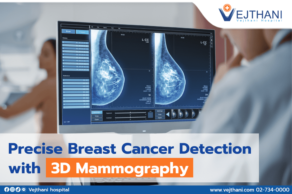
Mammography is an imaging technique that uses low-dose radiation to capture breast images. The radiation dose is slightly higher than chest X-ray, roughly one-sixth of the natural radiation in the atmosphere. Vejthani Hospital uses 3D mammography that takes only 3.7 seconds to capture each image for each angle, allowing clarity of overlapping breast tissues. This results in more detailed images, which is especially beneficial for patients with dense breast tissue. Annual mammograms are considered safe and effective in detecting breast abnormality in the early stage.
Ultrasound can reveal the presence of lumps or cysts in the breast. If a lump is detected, the ultrasound can determine its size and outline, helping to assess whether it is benign or more likely to be cancerous. This leads to a more precise diagnosis.
Breast cancer is a silent threat. Breast cancer screening with mammography and ultrasound is vital for all women with indicated risk factors. Early detection is key to successful treatment and cure.
For more information, please contact
Breast Center, Vejthani Hospital.
Call: (+66)2-734-0000 Ext. 2715
English Hotline: (+66)85-223-8888
- Readers Rating
- Rated 5 stars
5 / 5 ( Reviewers) - Spectacular
- Your Rating