Overview
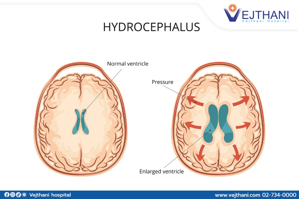
Hydrocephalus is a condition where there is an excessive buildup of cerebrospinal fluid (CSF) in the cavities (ventricles) deep inside the brain. The extra fluid makes the ventricles bigger and pressures the brain by expanding them.
Cerebrospinal fluid often circulates through ventricles in the brain. The brain’s CSF functions as a mechanism for delivering nutrients and removing waste. The brain and spinal cord are covered in CSF, which protects and cushions them from injury. However, the pressure of excessive CSF brought on by hydrocephalus can harm brain tissues and result in a variety of disorders with brain function.
The four types of hydrocephalus are as follows:
- Communicating hydrocephalus: Occurs when the circulation of cerebrospinal fluid (CSF) is obstructed after it exits the ventricles. This particular form of hydrocephalus may arise from the thickening of membranes known as arachnoid, located at the base of the brain. The thickened arachnoid membranes impede the unobstructed passage of CSF, leading to the condition. It is referred to as “communicating” because the CSF can still move between the ventricles, which remain unobstructed.
- Non-communicating hydrocephalus: Which is also referred to as obstructive hydrocephalus, occurs when the movement of cerebrospinal fluid (CSF) is obstructed within the narrow channels that connect the ventricles.
- Normal pressure hydrocephalus (NPH): Occurs when cerebrospinal fluid (CSF) accumulates, leading to enlargement of the ventricles, but with minimal or no increase in pressure. Unlike other types of hydrocephalus, the key distinction of NPH is that the pressure within the ventricles remains unchanged, despite the presence of an excessive amount of CSF. The buildup of CSF in the ventricles happens gradually, and symptoms manifest gradually as well. NPH primarily affects the elderly population.
- Hydrocephalus ex-vacuo: Occurs as a consequence of brain injury or stroke, leading to damage in the brain tissue surrounding the ventricles. This damage causes a reduction in the size of the brain tissue, creating additional space within the ventricles. Consequently, cerebrospinal fluid (CSF) accumulates within the ventricles to compensate for the increased space. As a result, the ventricles become enlarged, while the pressure within the head typically remains within the normal range.
Although hydrocephalus can occur at any age, it is more common in infants and individuals who are 60 years or older. Normal levels of cerebrospinal fluid in the brain can be restored and maintained through hydrocephalus surgery. Various therapies are often required to address symptoms or issues related to hydrocephalus.
Symptoms
The symptoms of hydrocephalus can differ based on the individual’s age, the stage of the disease, and their ability to tolerate the accumulation of CSF.
Infants: Infants with hydrocephalus frequently exhibit the following symptoms:
- Changes in the head
- Unusually large head
- Abrupt increase in head size
- Enlarged or tensed soft area (fontanel) on top of the head.
- Nausea and vomiting
- Lethargic
- Irritability
- Eating poorly
- Seizures
- Sunken eyes
- Weakness and a lack of tone in the muscles
Toddlers and older children: Signs and symptoms in toddlers and older children may include:
- Abnormal enlargement of the head
- Headache
- Nausea or vomiting
- Vision problem
- Abnormal eye movements
- Abnormal enlargement of the head
- Lethargic
- Balance or gait problem
- Coordination problem
- Poor appetite
- Loss of bladder control or a need to urinate frequently.
- Irritability
- Change in personality
- Poor school performance
- Delays or issues with learned abilities, such walking or talking.
Young and middle-aged adults: For this age group, typical symptoms and indications include:
- Headache
- Sluggishness
- Coordination or balance problem
- Loss of bladder control or a need to urinate frequently.
- Vision problems
- A decline in memory, focus, and other cognitive abilities that could have an impact on job performance.
Older adults: The more common hydrocephalus indications and symptoms in people 60 years of age and older are:
- Loss of bladder control or a need to urinate frequently.
- Dementia or memory loss
- Progressive decline in other forms of reasoning or thinking.
- Coordination or balance problem
- Difficulty walking, which is frequently characterized by a shuffling stride or the sensation that the feet are trapped.
Infants and toddlers exhibiting high-pitched cries, feeding difficulties, sucking issues, unexplained vomiting, seizures, or other unexplained signs and symptoms should be urgently taken to the hospital. Prompt diagnosis and treatment are crucial as these issues could be indicative of hydrocephalus, which can be triggered by various illnesses. Ensuring immediate medical attention is essential to address the condition effectively.
Causes
Hydrocephalus occurs due to an imbalance in the production and absorption of cerebrospinal fluid (CSF). The ventricular tissues in the brain generate CSF, which then moves through interconnected channels within the ventricles. Eventually, the fluid spreads into the spaces surrounding the brain and spinal column. The primary absorption of CSF takes place through blood vessels present in the brain’s surface tissues. This disruption in the equilibrium between CSF production and absorption leads to the development of hydrocephalus.
One of the following causes can result in the ventricles having too much cerebrospinal fluid:
- Obstruction: The most typical issue is a partial obstruction of the cerebrospinal fluid flow, either from one ventricle to another or from the ventricles to other regions around the brain.
- Overproduction: Cerebrospinal fluid rarely develops faster than it can be absorbed.
- Poor absorption: Absorption issues with cerebrospinal fluid are less frequent. This is frequently linked to inflammation of the brain tissues as a result of illness or injury.
Any time after birth, acquired hydrocephalus can manifest itself and have an impact on people of all ages. the following are the primary factors that lead to acquired hydrocephalus:
- Head trauma
- Stroke
- Spinal cord or brain tumors
- Brain or spinal cord infection, such as meningitis.
Additionally, bleeding or post-operative problems could result in hydrocephalus with normal pressure. Without any apparent cause, NPH affects a lot of people.
Risk factors
The cause of hydrocephalus is frequently unknown. However, a number of medical or developmental issues can cause or contribute to hydrocephalus.
Newborns: Any of the following factors may result in congenital (existing at birth) or postpartum hydrocephalus:
- Unusual growth of the central nervous system that could impede cerebrospinal fluid flow.
- A potential consequence of premature birth is internal bleeding in the ventricles.
- An infection in the uterus during pregnancy, such as syphilis or rubella, which can lead to inflammation in the embryonic brain structures.
Other risk factors: That may cause hydrocephalus.
in people of any age include:
- Brain or spinal cord lesions or malignancies.
- Infections of the central nervous system, such as bacterial meningitis or measles.
- Brain bleeding as a result of a stroke or head injury.
- Other traumatic brain injuries.
Diagnosis
A neurological examination is used for diagnosing hydrocephalus. Typically, procedure used to make a diagnosis of hydrocephalus include:
- Neurological examination: Age will determine what kind of neurological examination is performed. The neurologist may asked questions and do quick tests in the clinic to assess the muscle tone, range of motion, general health, and sense of smell.
- Imaging tests: The following procedures may assist in the diagnosis of hydrocephalus and the determination of the root causes of the symptoms:
- Ultrasound: This procedure is commonly employed as an initial assessment for infants due to its simplicity and low-risk nature. During the test, an ultrasound device is placed over the fontanel, the soft spot on the baby’s head. Additionally, ultrasound can be utilized during routine prenatal examinations to potentially identify hydrocephalus before birth.
- Magnetic resonance imaging (MRI): A magnetic field and radio waves are used in this examination to provide precise photographs of the brain. Although this examination is painless, it is noisy, and one have to stay still. The enlarged ventricles brought on by too much cerebrospinal fluid can be seen on MRI scans. They can also be utilized to determine the underlying causes of hydrocephalus or other issues that may be causing the symptoms.
For some MRI scans, children may require some light sedation. However, certain medical facilities utilize a quick version of MRI that typically does not require any sedation.
-
- Computed tomography (CT) scan: Cross-sectional images of the brain are produced using this specialized X-ray technology. Scanning is rapid and painless. CT scanning exposes patients to a modest amount of radiation while producing less detailed images than MRI. Hydrocephalus CT scans are typically reserved for urgent cases. A child typically receives a slight sedative for this test because it also demands lying still.
Adults frequently have additional testing to diagnose the disease. These tests could consist of spinal tap or lumbar puncture, monitoring of the intracranial pressure, and fundoscopic exam that examine the optic nerve at the back of the eye.
Treatment
Currently, brain surgery is the only possible option for treating hydrocephalus. In order to treat hydrocephalus, there are two different kinds of brain operations:
- Shunt: The most common treatment for hydrocephalus involves a surgical procedure to implant a medical device known as a shunt. This device consists of a flexible tube with a valve that is placed in the brain to regulate the flow of cerebrospinal fluid (CSF). The excess CSF is redirected through the tube to another part of the body, such as the abdomen or a heart chamber, where it can be absorbed more efficiently. This drainage system helps alleviate the pressure and fluid buildup in the brain.
Individuals with hydrocephalus typically require a shunt system for the remainder of their lives. Regular monitoring is essential to ensure the shunt is functioning properly and to address any complications that may arise. This treatment approach enables the ongoing management of hydrocephalus by effectively controlling the fluid levels in the brain, allowing individuals to lead a more normal and comfortable life.
- Endoscopic third ventriculostomy (ETV): Is a surgical procedure primarily conducted on children over the age of 2, involving the creation of a small hole in the floor of the third ventricle. This opening serves as a pathway for cerebrospinal fluid (CSF) to circulate in and around the brain, restoring its normal flow. Using a miniature video camera, the surgeon gains visual access to the brain and carefully punctures either the base of one ventricle or the space between them, allowing the CSF to drain out of the brain and alleviate the underlying condition.
- Other treatments: Hydrocephalus can require additional treatment for individuals, especially children, depending on the severity of its long-term complications. In the case of school-going children, the presence of special education teachers becomes crucial. These educators play a vital role in addressing any learning disabilities, evaluating educational requirements, and identifying the necessary resources to support the child’s educational journey.
Similarly, adults facing more severe complications stemming from hydrocephalus may require the assistance of various professionals. Occupational therapists, social workers, specialists in dementia care, and other medical experts might be necessary to provide comprehensive support. These professionals cater to the specific needs of adults with hydrocephalus, helping them manage their condition and enhancing their overall well-being.


