Overview
Aortic valve disease is a disease that affect the valve between the body’s main artery (aorta) and left lower chamber of the heart (ventricle). The aortic valve controls blood flow by ensuring that it proceeds through the heart in the proper direction from the left ventricle to the aorta. Aortic valve disease can affect blood flow to the rest of the heart and body.
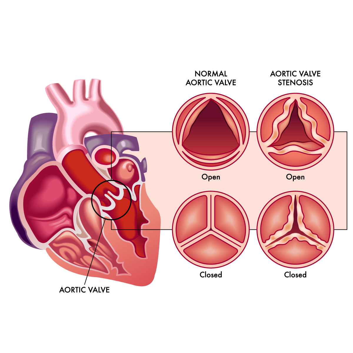
Types of aortic valve disease are as follows:
- Aortic valve stenosis: The aortic valve is thick and stiff, which causes the narrowing of the opening, which decreases or completely prevents blood flow to the body.
- Aortic valve regurgitation: Blood flows backward into the left ventricle as a result of an improperly closing aortic valve.
Aortic valve disease could occur from birth, or it could develop over time because of other medical conditions. Surgery and medications are the treatment options for aortic valve disease.
Symptoms
Aortic valve disease patients primarily as no symptoms but with time progresses to the following:
Aortic valve stenosis symptoms may include:
- Chest tightness or pain
- Rapid pulse rate
- Shortness of breath
- Fatigue
- Swelling of ankles
- Feeling dizzy or fainting
- Trouble with gaining weight (in children with aortic valve stenosis)
- Trouble with feeding (in children with aortic valve stenosis)
Aortic regurgitation symptoms may include:
- Chest tightness or pain
- Shortness of breath
- Fainting during excessive physical activity
- Palpitations
- Dizziness
- Swelling of ankles
If the patient has the signs and symptoms of aortic valve disease especially sudden chest pain, then emergency medical intervention is needed. Otherwise if you experience shortness of breath, exhaustion during exercise, or feelings of a pounding or irregular heartbeat, schedule an appointment with a medical professional.
Causes
Aortic valve disease can also develop as people age for the following reasons:
- Congenital heart defect: a defect in the structure of the heart that is present at birth
- Age-related changes to the heart: with increasing age, the valve will be thickening and loss of elasticity
- Infections in the heart valve, also called infective endocarditis
- High blood pressure
- Injury to the heart
Risk factors
Aortic valve disease risk factors include the following:
- Age: as people get older, calcium deposits can accumulate on the aortic valve, resulting in stiffening and narrowing of the aortic valve.
- Birth defect: congenital heart defects, patients who have congenital heart defect have higher risk to develop aortic valve regurgitation.
- Heart problem: an infection of the chambers’ lining and heart’s valves (endocarditis) may damage the aortic valve.
- Rheumatic fever: aortic stenosis, a form of valve disease, can be caused by strep throat complication. Rheumatic heart disease is the medical term for heart valve damage brought on by rheumatic fever.
- History of radiation therapy to the chest: patient who have history of radiation therapy have higher risk to develop aortic valve disease.
- Connective tissue disorder: Marfan syndrome, a connective tissue disorder can increase the risk or aortic valve disease.
- Chronic kidney disease: patient who have chronic kidney disease have an increased risk for the development of aortic valve disease.
Diagnosis
The diagnosis for both types of aortic valve disease is the same. A physical examination, questions about the signs and symptoms, and a medical history are typically used to determine whether the patient have aortic valve disease.
If during the assessment the specialist hear heart murmur (whooshing sound of the heart), the patient will be referred to a cardiologist.
Aortic valve disease can be detected using a number of tests, including:
- Echocardiogram: is a type of cardiac ultrasound which examines the aortic valve and aorta more closely is possible and determines the severity of aortic valve.
- Electrocardiogram (ECG or EKG): this procedure captures the heart’s electrical activity. Sensors (electrodes) are applied to the chest, wrists, and legs, then it is attached to the monitor to show the heart rhythm.
- Chest X-ray: is a noninvasive radiographic imaging that shows the heart and lungs. The test results may be used by a medical professional to determine whether the heart is enlarged, which may indicate certain forms of aortic valve disease or heart failure.
- Cardiac magnetic resonance imaging (MRI): this procedure produces precise pictures of the heart using magnetic fields and radio wave and helps to assess the degree of aortic valve disease and the size of the aorta.
- Cardiac computerized tomography (CT) scan: produces precise detailed pictures of the heart and heart valves using a set of X-rays, which helps to examine the aorta more carefully and determine its size. Aortic valve calcium levels, aortic valve stenosis severity, and aorta size may all be assessed with a CT scan.
- Exercise tests or stress tests: In these tests, an ECG or echocardiogram is frequently performed while the patient walks on a treadmill or on a stationary bike. Exercise tests can assist determine how the heart responds to exercise and whether exercising causes symptoms of valve disease.
- Cardiac catheterization: This test involves inserting a thin, flexible tube (catheter) into a blood vessel, typically in the arm or groin area, and directing it to the heart. Cardiac catheterization can provide greater information regarding blood flow and heart function. Cardiac catheterization can be used for some heart treatments.
Staging
The healthcare professional might describe the disease stage once testing confirms a diagnosis of aortic or another heart valve disease. The best suitable course of action is determined by staging.
The four main stages of heart valve disease are as follows:
- Stage A: Patient is at risk for heart valve disease. Categorize as “at risk”
- Stage B: Mild or moderate valve disease. There are no signs of cardiac valve disease. Categorize as “progressive”.
- Stage C: Although there are no symptoms associated with heart valve disease, the condition is severe. Categorize as “asymptomatic severe”.
- Stage D: Heart valve disease show signs and symptoms that could is in severe condition. Categorize as “symptomatic severe”.
Treatment
Treatment for aortic valve disease includes observation, dietary changes, medication, surgery, and other procedures. Someone with this disease should have a consultation and treatment with a cardiologist. Treatment will depend on the severity, staging, symptoms, and the condition of the patient.
Medications
Medication may be prescribed by the specialist if the aortic valve disease is mild to moderate or asymptomatic. Regular checkup is advised for close monitoring of the disease. Medications are used to treat underlying symptoms of the disease such as controlling blood pressure, preventing irregular heartbeats, removing any excess fluid from the body that could strain the heart or prevent further complications.
Surgery
The aortic valve condition may require surgical intervention or catheterization for repair or replacement regardless of the symptoms. Aortic valve surgery is frequently performed during an open-heart surgery. However, for some cases, a catheter-based method or minimally invasive cardiac surgery, which requires smaller incisions than open heart surgery, may be used to replace the valve. Discussing with the specialist the risk and benefits of each procedure could help the patient choose the best option.
- Aortic valve repair: is usually done with open-heart surgery to reconstruct form and function of an aortic valve. However, minimal invasive surgery may also be an option. A less invasive technique called balloon valvuloplasty may be used in newborns and kids with aortic valve stenosis to open a stenotic heart valve. To enlarge the valve opening, a balloon is inflated, deflated, and then removed. Adults who are too severely ill for surgery or who are waiting for a valve replacement may also undergo this valve repair technique.
- Aortic valve replacement: An aortic valve replacement is usually performed through an open-heart valve surgery to remove damage valve and replace the heart valve with a new valve whether synthetic or animal tissue valve (biological valve). In some cases, the Ross procedure is used to treat aortic valve disease by remove the damaged aortic valve and replace with it with pulmonary valve.
- Transcatheter aortic valve replacement (TAVR): is a minimally invasive surgery, uses to replace a constricted aortic valve with a biological tissue valve. Compared to open heart surgery, TAVR will requires smaller incisions. For those who are more vulnerable to complications following cardiac valve surgery, TAVR may be a better option.
The specialist will discuss all of your treatment option with the risk and benefit of each type of valve and procedure to choose the appropriate treatment for you.