Overview
Aneurysm is the development of an abnormal bulge or ballooning at the blood vessel’s wall. Aneurysm occur when they are weakening of the blood vessel wall or a weakness in the artery’s wall which could balloon outward due to the pressure of the blood passing through it.
A ruptured aneurysm can cause fatal bleeding. Aneurysms may not cause symptoms even if they have grown bigger, causing the patient not to be aware of their condition.
The following are types of aneurysms that could develop within the body:
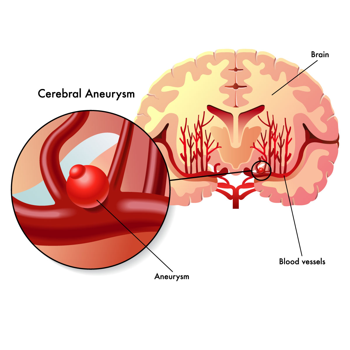
- Brain aneurysm (cerebral aneurysm): The blood vessels that supply blood to the brain are affected.
- Aortic aneurysm: The body’s aorta, which transports blood to the body’s other organs from the heart, is affected.
- Abdominal aortic aneurysm: The aorta portion that travels through your abdomen is affected.
- Thoracic aortic aneurysm: The aorta portion that travels through the chest (upper portion of the abdomen) is affected.
- Peripheral aneurysm: affects the blood vessels in other parts of your body, like your legs, groin, or neck.
- Carotid aneurysm: A rare type of aneurysm where blood vessels that deliver blood to the brain, neck, and face are affected.
To determine the possibility of the aneurysm rupture, the specialist will perform full physical examination, evaluate the size, location, and the appearance of the aneurysm. Small aneurysms may show very minimal risk of rupture; however, specialist will monitor and determine if further treatment such as surgery is needed.
Classification of aneurysm:
Classification if based on the location, size, and the appearance of the aneurysm:
- Fusiform aneurysm: the bulging outward sides of the arteries.
- Saccular aneurysm: the bulge is located only at one side for the artery.
- Mycotic aneurysm: an infection was developed at the valves of the heart and cause arterial wall to weaken.
- Pseudoaneurysm or false aneurysm: The outer layer of the arterial wall may expand as a result of damage to the artery’s inner layer.
Symptoms
Aneurysm is a life-threatening condition, therefore if any signs or symptoms are seen, it is important to call the healthcare provider. The following are the symptoms of ruptured aneurysm:
- Lightheadedness.
- Fatigue
- Nausea and vomiting
- Difficulty in swallowing
- Palpitation or rapid heartbeat.
- Sudden severe headache
- Sudden chest pain
- Sudden abdominal pain, or back pain.
- Sudden drop of blood pressure
- Loss of consciousness
- Confusion or dizziness
Cause
Aneurysms can occasionally develop at birth and may also appear at any time during life. Although the exact cause of an aneurysm is unknown, several potential explanations include:
- Atherosclerosis: blockage inside the walls of the arteries that cause by a buildup plaque.
- Family history of aneurysms
- High blood pressure: which can damage and weaken the aortic walls.
- Deep wound infections: can also lead to aneurysm.
Risk factors
There are many risk factors depending on the types of aneurysms as follows:
- Gender: male commonly have abdominal aneurysm and aortic aneurysm, while female have brain aneurysms.
- Age: an aneurysm are mostly found in people who are over the age of 60.
- Smoker: people who smoke have higher risk to develop abdominal aneurysm.
- Race: Non-African American people have higher risk of developing the disease.
Diagnose
Aneurysms may occur without signs and symptoms, and there are cases the aneurysm was discovered by the specialist when conducting assessment or by other procedures.
A specialist will conduct imaging test if the patient have any symptoms that could point to an aneurysm. The following procedures or test may help the specialist detect and diagnose an aneurysm:
- Computed tomography (CT) scan: which is painless as it uses X-rays to create cross-sectional images of the body structures as well as clear images of the vessel. This helps to determine the size and shape of an aneurysm. While the doctor is performing a CT scan, the doctor will ask you to lie on a table which slides into the scanning machine. The doctor might also inject contrast material into a vein to make your blood vessels show up more clearly on the images.
- Magnetic resonance imaging (MRI) angiogram: a diagnostic technique called an angiography uses imaging to demonstrate how blood flows through your heart or blood vessels. A contrast is then injected to help visualize the blood flow and show the location of the blockage.
- Ultrasound: by using sound waves to create real time images of the internal organ, including the vessel. A technician will perform an ultrasound by putting a slight pressure on an ultrasound probe (transducer) on the area of the body.
Treatment
Aneurysms are treated to prevent aneurysms from rupturing through medication and surgery. The doctor will select the treatment based on the aneurysm’s size and progression of the disease.
Medication
Medication helps reducing blood pressure, enhancing blood flow, and managing cholesterol levels, all of which could decrease the growth of aneurysms and reduce pressure on the arterial wall.
Surgery
- Open surgery: An incision is made to conduct the graft or remove the aneurysm.
- Endovascular aneurysm repair: A catheter (thin tube) is inserted into the vessel during endovascular surgery. The surgeon places a graft (a piece of specialized tubing) into the catheter to strengthen or repair the artery.
- Thoracic endovascular aortic repair (TEVAR): is a procedure to treat an aneurysm in the chest area.
- Fenestrated endovascular aortic aneurysm repair (FEVAR): is a procedure to treat a complex aortic aneurysm.
- Endovascular coiling: Is a procedure used for aneurysm occurring in the brain. The surgeon will inserts several coils by using a platinum wire spiral to block the blood flow into an aneurysm and will reduce risk of rupture aneurysm.
- Microvascular clipping: Is a procedure that cut off blood flow to the brain aneurysm by using a metal clip. Microvascular clipping will prevent aneurysm from rupturing.
- Catheter embolization: Through this surgery, the aneurysm’s blood supply is shut off. In order to administer medication or embolic agents that stop bleeding, the surgeon inserts a catheter into the injured artery.


