Aortic valve regurgitation
Overview
Aortic valve regurgitation, also known as aortic regurgitation, is a condition that develops when the leaflets known as cusps which open and close in a valve are not closed as tightly as they should, causing blood flow that is pumped out of the left ventricle (the main chamber of the heart) leak back into the chamber as a result of the disease. The leakage of aortic valve regurgitation prevents the heart from functioning properly by pumping blood to the rest of the body, which results in fatigue, or tiredness, or shortness of breath. This disease could suddenly develop, or it may take years to progress.
However, if it becomes severe surgical intervention is needed to repair or replace the aortic valve.
Symptoms
As aortic valve regurgitation is a slowly progressive disease, patients may be unaware of the disease as there may be no signs and symptoms. In some cases, the disease occurs suddenly due to a valve infection.
The signs and symptoms may include:
- Shortness of breath
- Chest pain, discomfort or tightness
- Fatigue
- Heart murmur
- Irregular pulse (arrhythmia)
- Lightheadedness or fainting
- Rapid heartbeat or palpitation
- Swelling of ankles and feet
If the patient is having any signs and symptoms, please consult the doctor.
Heart failure may occasionally be a factor in the development of aortic valve regurgitation. It is recommended to have a physical consultation with the specialist if the patient is experiencing tiredness that doesn’t improve with rest, shortness of breath, and swelling of ankles and feet, this may indicate heart failure.
Causes
One of the four valves that regulates blood flow through the heart is the aortic valve. It separates the major artery supplying your body with oxygen-rich blood (aorta) from the left ventricle, the heart’s primary pumping chamber. Each heartbeat causes the valve’s flaps (also known as cusps or leaflets) to open and close once. Aortic valve regurgitation develops when the blood flows regurgitate back into the left ventricle as a result of an abnormal closing of the aortic valve and it can lead to an enlargement of the heart chambers.
The larger left ventricle essentially exerts more force to keep a regular blood flow. Due to the disease the left ventricle and the heart eventually become weaker. Aortic valve regurgitation is a slow progressive disease (chronic) but it can induce instantly (acute) as a result of valve infection.
The following are some causes of aortic valve regurgitation:
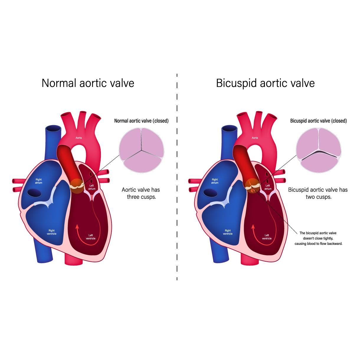
- Congenital heart valve disease: These are at risk of experiencing aortic valve regurgitation at some point in their life because of these congenital heart defects. Rather than typical three distinct cusps valves, some people are born with aortic valves that have two cusps (bicuspid valve). In rare cases unicuspid and quadricuspid valves can occasionally exist.
A bicuspid valve increases the risk of developing one if they have a parent or sibling who has the disease. Even without a history of the disease in the family, someone can still have a bicuspid valve.
- Aortic stenosis (Narrowing of the aortic valve): this condition prevents the valve to open or close properly because of the blockage. This may be due to the calcium deposits that was build up that aortic valve stiffening and narrowing.
- Endocarditis: Inflammation of the lining of the heart’s chambers and valves. Infection is typically the cause of this life-threatening condition. The aortic valve may be damaged by it.
- Rheumatic fever: The damage to the heart valves might result from untreated strep throat infection. The aortic valve may develop scar tissue that will make the opening of the aortic valve become narrow, or it may become rough, allowing calcium deposits to build up. It’s called rheumatic heart disease.
- Other diseases: Marfan syndrome, a connective tissue disorder, and some autoimmune diseases such as lupus can cause the aorta and aortic valve to expand and cause regurgitation.
- Tear or injury at the aorta: blood may flow backward through the aortic valve as a result of a traumatic chest injury or an aortic tear (dissection).
Risk factors
The following are some risk factors that will likely develop into aortic valve regurgitation:
- Age: elderly is more like to be affected by the disease.
- Congenital heart disease: some certain heart conditions from birth could lead to aortic valve regurgitation.
- Previous infection: past illnesses such as rheumatic fever and infective endocarditis may have damaged the heart
- Other conditions: Marfan syndrome and aortic valve stenosis that can affect the heart.
- High blood pressure: patient who have high blood pressure have an increased risk of the developing aortic valve regurgitation.
Diagnosis
Aortic valve regurgitation could be diagnosed during a physical examination, assessment, and medical history. If during the assessment the specialist detected heart murmur (whooshing sound of the heart), the patient will be referred to a cardiologist.
Aortic valve regurgitation can be diagnosed through the following tests:
- Imaging:
- Chest X-ray: the heart can be determined through a chest X-ray. It can also assist medical professionals in assessing the condition of the lung as well as revealing aortic enlargement and calcium deposit on the aortic valve.
- Cardiac magnetic resonance imaging (MRI): This test creates precise images of your heart using magnetic field and radio wave and help to assess the size of the aorta and the degree of aortic valve regurgitation.
- Stress test or exercise tests: In these tests, an ECG or echocardiogram is frequently performed while the patient walks on a treadmill or on a stationary bike. Exercise test can assist to determine how the heart responds to exercise and whether exercising will trigger development of the aortic valve symptoms. If the patient is unable to exercise, they may be able to benefit from using medications that have the same heart-healthy effects as exercise.
- Echocardiogram: Images of the beating heart are produced by sound waves that being sent by the heart using a transducer, that is held on the chest. The test assesses the severity, the cause and the condition of the aorta and the aortic valve.
The aortic valve may be examined more closely using a transesophageal echocardiography which involves inserting a small transducer end tube from the mouth and down to your esophagus.
- Electrocardiogram (ECG or EKG): this procedure captures the heart’s electrical activity. Sensors (electrodes) are applied to the body on the chest, wrists, and legs, then it is attached to the monitor to show the heart rate. An ECG can also identify cardiac disease and enlarged heart chambers.
- Cardiac catheterization: In some cases that the other tests are not suitable for diagnosis and severity, this test can be used for diagnosis of aortic valve regurgitation.
Prior to aortic valve surgery, this test is used to evaluate coronary artery blockages.
The procedure will insert a narrow tube (catheter) into a blood vessel, commonly at the wrist or groin area, and then into the heart. A dye runs through the catheter to improve the visualization of the arteries on X-rays, this procedure called a coronary angiogram.
Treatment
Treatment for aortic valve regurgitation is used to reduce symptoms and prevent complications. The choice of treatment depends on severity of the disease. Specialist may propose a healthy lifestyle modification and schedule a routine follow-up to closely monitor the condition if the patient’s symptoms are mild or the patients aren’t having symptoms. Patients may need regular echocardiograms to prevent worsening of the condition.
Medications
Medications will be prescribed to treat the symptoms or prevent complications of aortic valve regurgitation.
Surgery
If the patient has severe aortic regurgitation and symptoms, they may require surgery to repair or replace the damaged aortic valve. However, even when the condition is mild or there are no symptoms, a person may also need the surgery.
Aortic valve repair or replacement may be performed as an open-heart procedure that requires a chest incision, or it may be performed using a minimally invasive heart surgery to replace the damaged aortic valve, smaller incisions are made during these procedures like transcatheter aortic valve replacement (TAVR) than during open-heart surgery.
Whether an aortic valve replacement or repair is necessary, it will depend on the symptoms, age, overall health, and whether the patient also require a heart surgery to correct another heart problem.
Surgical options for aortic valve regurgitation includes:
- Aortic valve repair: Separating the fused valve flaps (cusps), reshaping or removing any extra valve tissue to allow the cusps to shut firmly, or repairing valve holes are the methods used in this treatment. In order to fix a replacement aortic valve that is leaking, doctors may perform a catheter procedure.
- Aortic valve replacement. An aortic valve replacement is usually performed through an open-heart valve surgery to remove damage valve and replace the heart valve with a new valve whether synthetic or biological valve (animal tissue valve).
Transcatheter aortic valve replacement (TAVR) is a minimally invasive treatment to replace an aortic valve with a biological tissue valve. Compared to open heart surgery, TAVR will require smaller incisions. For those who are more vulnerable to complications following cardiac valve surgery, TAVR may be a better option.
In some cases, the Ross procedure is used to treat aortic valve disease by removing the damaged aortic valve and replace with it with pulmonary valve.
Biological tissue valves deteriorate over time and may eventually require replacement. To prevent blood clots, people with mechanical valves must take blood-thinning drugs continuously. The specialist will discuss all of your treatment option with the risk and benefit of each type of treatment options and procedure to choose the appropriate treatment for the patient.
Lifestyle and home remedies
Doctor will check on with the patient on a regular basis at follow-up appointments. These heart-healthy habits will help preventing or slowing the progression of heart disease.
- Consuming heart-healthy diet: Consume a range of fruits and vegetables, dairy products with low fat or no fat, chicken, fish, and whole grains. Steer clear of extra salt, sugar, and saturated and trans fats.
- A healthy weight: Maintain a healthy weight. If the patient is obese or overweight, the doctor could advise them to lose weight.
- Regular physical activity: As a regular part of your fitness program, aim to get in around 30 minutes of exercise. However, before beginning an exercise regimen, especially if the patient would like to participate with competitive activity, they might need to get doctor’s approval.
- Stress management: The patient may need to manage their stress with restorative exercises, meditation, physical activity, and spending quality time with loved ones.
- Avoiding tobacco: Avoid smoking. Discover tools to assist you in quitting smoking by speaking with your doctor.
- Controlling high blood pressure. Follow your doctor’s instructions completely while using any blood pressure medicine.


